|
When people see pictures of bodies, a whole range of
brain regions are active. This network is altered in
women with anorexia nervosa.
In a functional magnetic resonance imaging study,
two regions that are important for the processing of
body images were functionally more weakly connected
in anorexic women than in healthy women. The
stronger this "connection error" was, the more
overweight the respondents considered themselves.
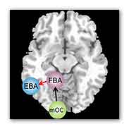
PHOTO - Network for body processing: When people
look at body images, the visual information first
enters in the central occipital lobe (mOC). The
“fusiform body area” (FBA) and the “extrastriate
body area” (EBA) then process the images further. In
anorexic women, the connection from the mOC to the
FBA is unaffected (black arrow). However, the FBA
and EBA do not work normally together in the left
hemisphere (red arrow). Credit: Boris Suchan.
"These alterations in the brain could explain why
women with anorexia perceive themselves as fatter,
even though they are objectively underweight" says
Prof. Dr. Boris Suchan of the Institute of Cognitive
Neuroscience at the Ruhr-Universität.
Together with Prof. Dr. Dietrich Grönemeyer
(University of Witten-Herdecke), Prof. Dr. Silja
Vocks (University of Osnabrück) and other colleagues,
the Bochum researchers report in the journal
Behavioural Brain Research.
The researchers tested ten anorexic and fifteen
healthy women of similar age. To start with, all the
women judged on the computer which of several
different silhouettes corresponded best to their own
body shape.
Ten control subjects who did not participate in the
MRI scan answered the same question by matching a
photo of the test subject to the right silhouette.
Both healthy and anorexic women estimated their body
shape differently than outsiders: healthy subjects
rated themselves as thinner than the control
subjects. Anorexic women on the other hand perceived
themselves to be fatter than the control subjects
did.
In MRI scanners, the researchers then recorded the
brain activity of the 25 participants while they
observed photos of bodies.
Above all, they analysed the activity in the "fusiform
body area" (FBA) and the "extrastriate body area" (EBA),
because previous studies showed that these brain
regions are critical for the perception of bodies.
To this end, the neuroscientists from Bochum
calculated the so-called effective connectivity
between the FBA and EBA in both hemispheres. This is
a measure of how much the activity in several brain
areas is temporally correlated. A high degree of
correlation is indicative of a strong connection.
The connection between the FBA and EBA was weaker in
women with anorexia nervosa than in healthy women.
In addition, the researchers found a negative
correlation between the EBA-FBA connection in the
left hemisphere and the misjudgement of body weight:
the weaker the effective connectivity between the
EBA and FBA was, the fatter the subjects with
anorexia falsely estimated themselves to be. "In a
previous study we found that there are structural
changes in the brains of patients with anorexia",
says Boris Suchan. They have a lower density of
nerve cells in the EBA. "The new data shows that the
network for body processing is also functionally
altered." The EBA, which has a lower cell density in
anorexics, is also the area that stood out in the
connection analysis: it receives reduced input from
the FBA. "These changes could provide a mechanism
for the development of anorexia", says Suchan.
More information
Suchan, B. et al. Reduced connectivity between the
left fusiform body area and the extrastriate body
area in anorexia nervosa is associated with body
image distortion.
http://www.sciencedirect.com/science/article/pii/S0166432812007760
http://www.ruhr-uni-bochum.de
(MDN)
|