|
During REM sleep, the brain inhibits the motor
system, which makes the sleeper completely immobile.
CNRS researchers working in the Centre de Recherche
en Neurosciences de Lyon (CNRS/Université Claude
Bernard Lyon 1/INSERM/Université Jean Monnet) have
identified a population of neurons that is
responsible for this transient muscle paralysis.
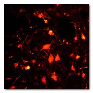
The glutamate neurons of the sublaterodorsal nucleus
emit a spontaneous red fluorescence indicating that
the viral vectors used have been successfully added.
© Sara Valencia Garcia / Patrice Fort, CNRS
The animal model created will shed light on the
origin of some paradoxical sleep disorders, and more
particularly the condition that prevents this
paralysis. It will also be most useful in the study
of Parkinson’s disease, since these pathologies are
related. This work was published on December 12,
2016 on the website of the journal Brain.
In spite of being in a deep sleep, the patients
talk, move, kick and eventually fall out of bed.
They are suffering from a parasomnia called REM
Sleep Behavior Disorder[1] (RBD).
This disorder usually appears around the age of 50.
Muscles are at rest during the REM sleep phase, but
in these patients, there is no paralysis, although
the reason for this is not known. The sleepers move
abnormally, probably reflecting their dream
activity.
A team from the Centre de Recherche en Neurosciences
de Lyon (CNRS/INSERM/Université Claude Bernard Lyon
1/Université Jean Monnet) has taken one more step
towards elucidating this pathology.
The researchers identified neurons in the
sublaterodorsal nucleus of the brain, ideally
located to control motor system paralysis during REM
sleep. In rats, they specifically targeted this
neuron population, by adding genetically modified
viral vectors to it.
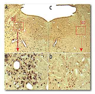
In a normal rat specimen (A and B) the neurons of
the sublaterodorsal nucleus (SLD, colored in brown)
are glutamate neurons (also colored in black). In
rats treated with viral vectors (C and D), neurons
are still present (in brown) but are not longer
capable of releasing glutamate (absence of black
color). © Sara Valencia Garcia / Patrice Fort, CNRS
Once these are in the neural cells, they block the
expression of a gene that allows synaptic glutamate
secretion. Now incapable of releasing this
excitatory neurotransmitter, the neurons can no
longer communicate with their neighbors. They are
disconnected from the cerebral network necessary for
paralysis during REM sleep.
For 50 years, the scientific community has
considered that these glutamate neurons generated
REM itself.
This team’s experience invalidates this hypothesis:
despite the absence of activity in this neuron
circuit, the rats still experience this stage of
sleep.
They are fast asleep and disconnected from the
outside world, with eyes closed.
But these rats are no longer paralyzed.
Their behavior is very reminiscent of the clinical
profile of patients suffering from RBD. The
glutamate neurons targeted in this study play an
essential part in REM paralysis during sleep and are
reportedly the first neurons affected in this
neurological disease.
This research work goes beyond creating a new
preclinical model that mimics this parasomnia. It
may be of paramount importance in studying some
neurodegenerative diseases.
Recent clinical research has shown that patients
diagnosed with RBD almost always develop the motor
symptoms of Parkinson’s disease, on average a decade
later.
The team is now attempting to develop an animal
model that evolves from parasomnia into Parkinson’s
disease, in order to understand how neuron
degeneration occurs.
See also
Sleep Paralysis: symptoms, causes and treatment
(2016-04-03)
Link...
For more information
Brain
A Journal of Neurology
Genetic inactivation of glutamate neurons in the rat
sublaterodorsal tegmental nucleus recapitulates REM
sleep behaviour disorder
Link...
Inserm
Institut national de la santé et de la recherche
Link...
Le Centre national de la recherche scientifique -
CNRS
Link...
MDN |